
Have you ever wanted to see what the world looks like at a microscopic level? With photomicrography, you can do just that! This process uses a microscope and a camera to capture images of small objects or features that are too small for the naked eye to see. These images can show us the intricate structure and behavior of cells, tissues, and other tiny structures, giving scientists and researchers a better understanding of the world around us.
But photomicrography isn’t just for scientists. Anyone with a curious mind and a sense of adventure can get in on the action. Imagine peering through a microscope and discovering a whole new world full of colorful cells, delicate tissues, and intricate patterns that you’ve never seen before. Each image is like a window into a different dimension, full of endless possibilities for discovery.
The Nikon Small World Competition is an annual photomicrography competition that showcases the beauty and complexity of the microscopic world. Photomicrographs (images taken through a microscope) of any microscopic subject matter can be submitted to the competition. The winning images are chosen based on their scientific merit, beauty, and originality. The competition has been held annually since 1975 and is open to anyone who has taken an image using microscopy equipment. Winning images are chosen by a panel of judges and are showcased in an online gallery as well as in a traveling exhibition. The competition is sponsored by Nikon Instruments Inc., a division of Nikon Corporation.
So, here are 20 of the winning and honored images of our own picks for 2022:
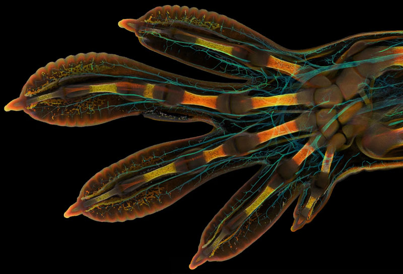
1. First Place Winner: Embryonic hand of a Madagascar giant day gecko (Phelsuma grandis)
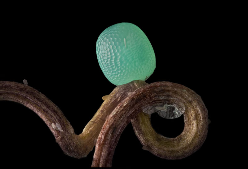
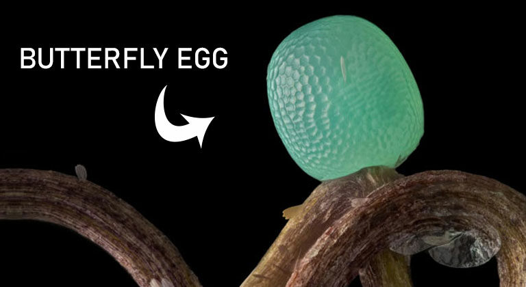
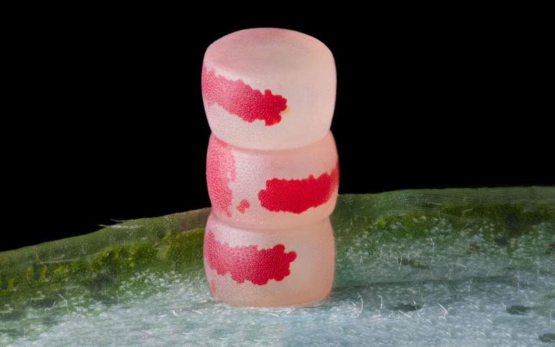
2. Honorable Mention: Butterfly egg
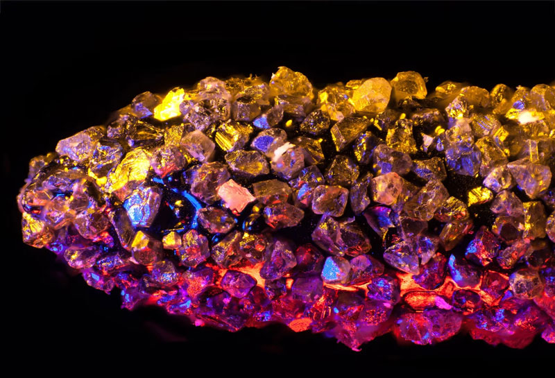
3. Image of Distinction: A dental drill bit studded with diamond chips
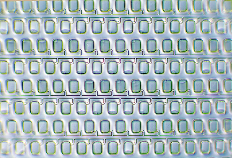
4. Image of Distinction: A digital PCR plate set up with RNA extracted from viruses in wastewater sludge
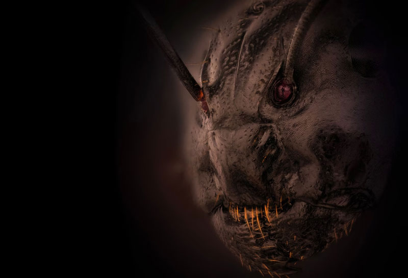
5. Image of Distinction: A close view of an ant (Camponotus)
6. Image of Distinction: Amino acid crystals (L-glutamine and beta-alanine)
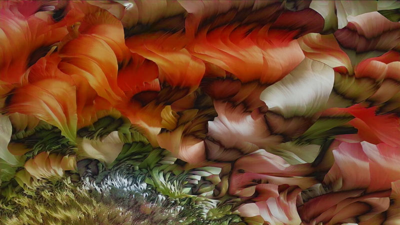
7. Image of Distinction: Four o’clock flower (Mirabilis jalapa)
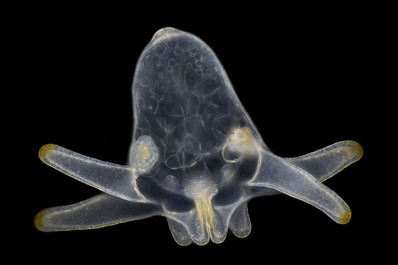
8. Honorable Mention: Larva of an anemone, found in marine plankton

9. Image of Distinction: Drops of olive oil in water
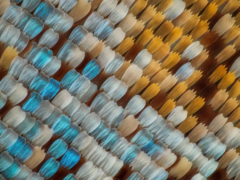
10. Image of Distinction: Butterfly scale
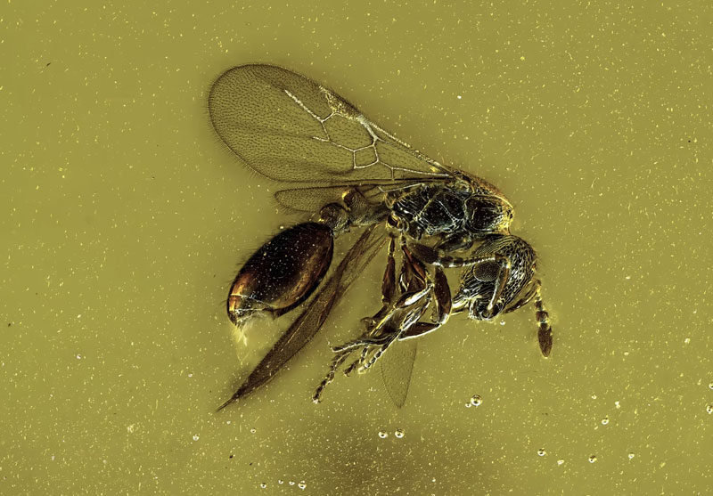
11. Image of Distinction: Winged ant encased in approximately 20-million-year-old Dominican amber
12. Image of Distinction: Moss spore capsule (sporangium)
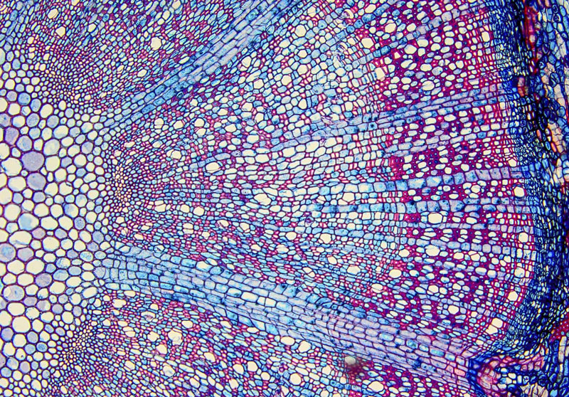
13. Image of Distinction: Wood cells
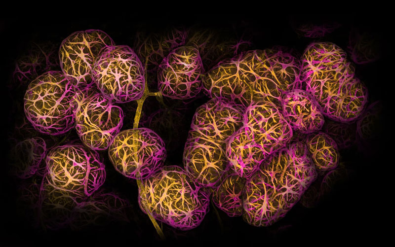
14. 11th Place Winner: Moth eggs
15. 2nd Place Winner: Breast tissue showing contractile myoepithelial cells wrapped around milk-producing alveoli
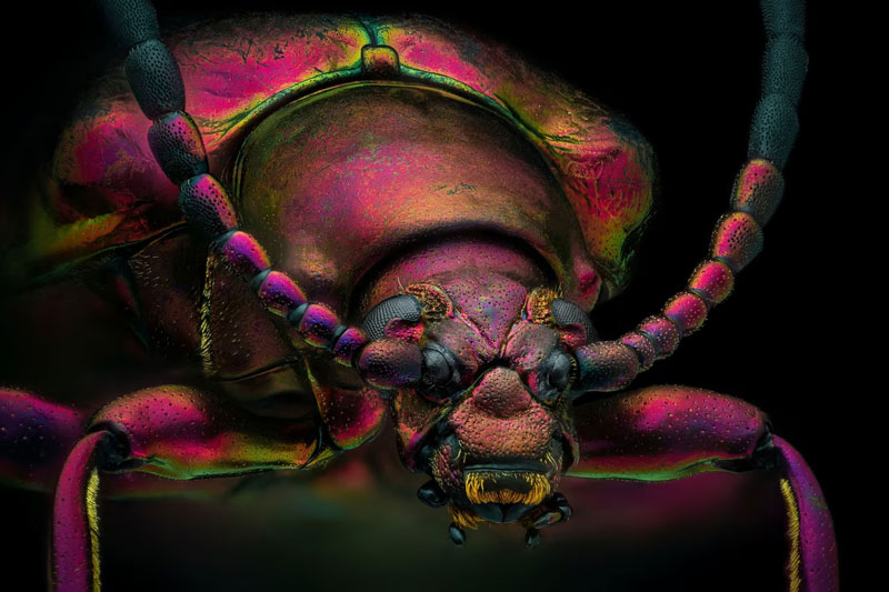
16. Image of Distinction: Red speckled jewel beetle (Chrysochroa buqueti rugicollis)
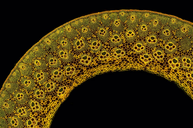
17. Honorable Mention: Young stem of garden bamboo (Fargesia sp.)
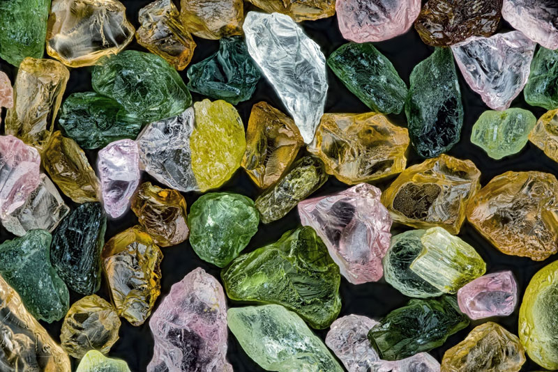
18. Image of Distinction: Grains of sand from Alaska
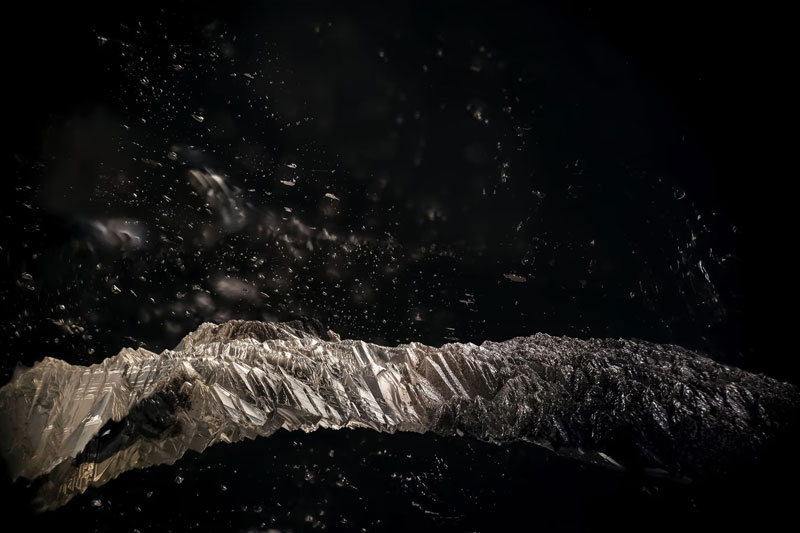
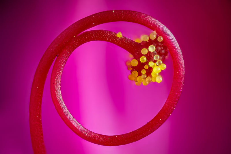
19. Image of Distinction: Etch tube in Brazilian quartz with iron oxide staining
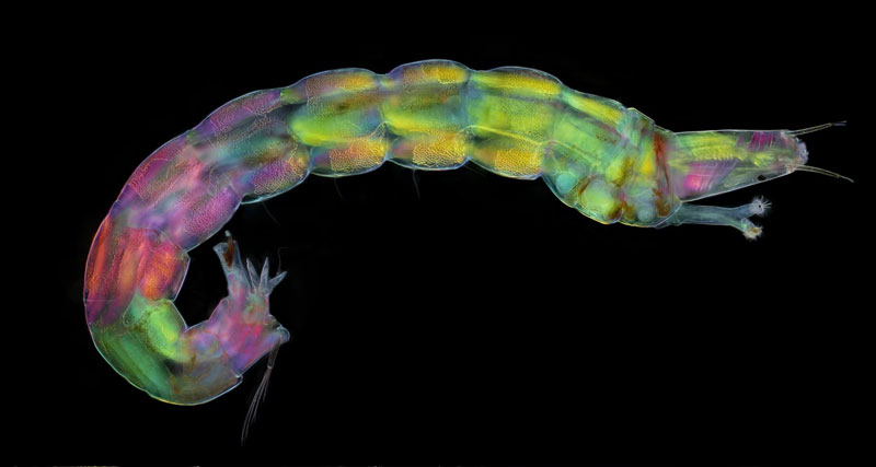
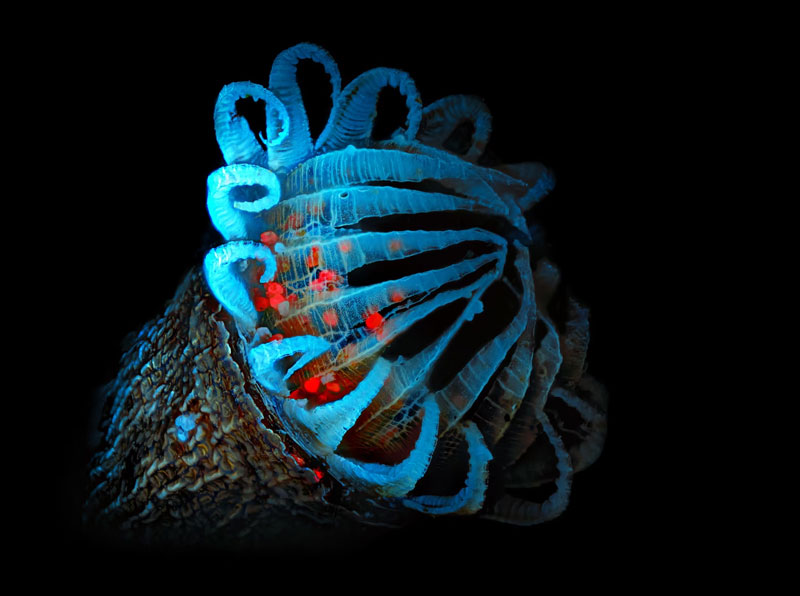
20. Honorable Mention: Midge larva collected from a fresh water pond